No products
Product successfully added to your shopping cart
There are 0 items in your cart. There is 1 item in your cart.


AJ-6340 Ophthalmic Ultrasound A/B Scanner Full Digital Touch Screen
Ophtamologie
- Consommables
- Equipements
- Mobiliers
- Protection
- Hygiène
Manufacturers
Viewed products
-

AJ-6340 Ophthalmic...
Ophthalmic Ultrasound A/B Scanner...


AJ-6340 Ophthalmic Ultrasound A/B Scanner Full Digital Touch Screen
New product
Ophthalmic Ultrasound A/B Scanner AJ-6340 with normal, vitreous bod enhancement, retina observation mode, mainly used for diagnosis of intraocular diseases, display the location, shape range of the focus of infection and the relationship with the surrounding tissue. Can be diagnosed vitreous opacity, retinal detachment, eye base tumors etc. eye diseases.
By buying this product you can collect up to 91440 loyalty points. Your cart will total 91440 points that can be converted into a voucher of 9 144 F.
- Write a review
- Send to a friend
- Remove this product from my favorite's list.
- Add this product to my list of favorites.

More info
-
Ophthalmic Ultrasound A/B Scanner AJ-6340 with normal, vitreous body enhancement, retina observation mode, mainly used for diagnosis of intraocular diseases, display the location, shape range of the focus of infection and the relationship with the surrounding tissue. Can be diagnosed vitreous opacity, retinal detachment, eye base tumors etc. eye diseases. A scan is used to measure anterior chamber depth, lens thickness, axial length, calculate diopter of implant IOL as well.
Ophthalmic Ultrasound A/B Scanner AJ-6340 works with PC workstation to compose a complete A/B scan system to support all the functions need to obtain during examinations:
- Measurements of the axial length (AL), anterior chamber depth(ACD), and lens thickness of an eye and for calculating the associated IOL power for an implanted lens
- View of the images of lens, vitreous, retina, choroid and sclera
- Statistic analysis of the obtained images
- Editable clinical report with all needed information
- Huge saved database capability
- Saved cases review
SPECIFICATIONS
B Scan
Frequency: 10MHz/20MHz (optional) ,Magnetic driven, noiseless
Scanning Mode: Sector Scanning
Magnify:Multi continuous magnification,Real-Time magnification
Resolution: Lateral ≤0.3mm; Vertical≤0.2mm
Geometry position precision: Lateral ≤10%; Vertical≤5%
Depth:60mm
Enhance the part of vitreous body and retina
Gain of probe:30dB-105dB
Scanning Angle:53°
Gray Scale: 256
False Color: Multi colors. OCT
measurement type: multigroup distances, perimeters and areas
Image postprocessing: multiple curves processing, Pseudo-color processing curve
Movies: 100 images movie review, AVI JPG format image output
A Scan
Frequency:10MHz, with LED
Depth: 40mm
Precision:±0.05 mm
Measurement: Anterior chamber depth, lens thickness, vitreous body length, total length and average
Eye mode: Phakic / Aphakic / Dense / Various IOL
IOL Formula: SRK-II, SRK-T, HOFFER-Q, HOLLADAY,BINKHORST-II, HAIGIS
Stat. Calculation: Average and standard deviation
Store: 10 Scanning results for each eye
Others
Display Mode :B, B+B, B+A, A
Hint: preset keyword
Case Search:Multi-keywords
Screen: 15 inch LCD
Built-in battery: can use 4 hours
User-defined report template poor credit secured loans
Reviews
24 autres produits dans la même categorie:
-

AJ-5D / 5D1 / 5D2 Microscope...
3 348 000 F
-

AJ-5E Microscope à lampe à...
2 970 001 F
-

AJ-5P Microscope à lampe à...
2 790 004 F
-

AJ-5S Microscope à lampe à fente...
2 700 005 F
-

AJ8B & AJ6B Trousse de...
234 000 F
-

AJ8C (DC.) Ophtalmoscope
147 600 F
-
AJ8A (alimenté) Ophtalmoscope
234 000 F
-
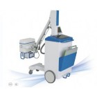
AJ-8000 Appareil de radiographie...
93 060 000 F
-

AJ-6800 Réfractomètre automatique
4 383 000 F
-

YZ30 Tonomètre
684 000 F
-

AJ-6400 Émulsifiant Phaco
22 499 999 F
-

Auto-réfracteur Huvitz HRK-7000
10 053 800 F
-

AJ-099 Ophthalmology O.T. Table
1 868 589 F
-

AJ-2000B Electric Ophthalmology...
3 929 400 F
-

Périmètre de projection
8 640 000 F
-

Périmètre de projection
11 448 000 F
-

Tonomètre sans contact avec...
11 035 596 F
-

Tonomètre sans contact
8 404 172 F
-

AJ-6300 Ophthalmic...
4 608 000 F
-

Ophalmoscope HS-OP 10C
208 860 F
-

Negato. 1plage (MT01002021)
153 400 F
-

Negato. 2 plage (MT01002022)
188 800 F
-

Ophtalmoscope
177 000 F
-

Ensemble ophtalmoscope d'otoscope
53 100 F

support service
Lorem ipsum dolor sit amet conse ctetur voluptate velit esse cillum dolo

free delivery
Lorem ipsum dolor sit amet conse ctetur voluptate velit esse cillum dolo

secure payment
Lorem ipsum dolor sit amet conse ctetur voluptate velit esse cillum dolo
© 2015 ChoiceMarket Demo Store. All Rights Reserved.


